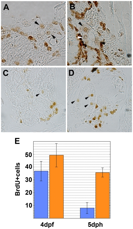
Figure 7. Cell proliferation in the renal tubules and ducts.
Cell proliferation in the kidney was compared between (A, C) wild-type and (B, D) pc mutant medaka at 4-day postfertilization (dpf) (A, B) and 5-day posthatching (dph)(C, D). Arrowheads indicate BrdU-positive cells in the renal ductal or tubular epithelia. (E) Number of BrdU-positive cells in the epithelium of the anterior portion (most anterior 15 sections) of the pronephros. Sections were obtained from four wild-type and four pc mutant fry at both 4 dpf and at 5 dph. The number of BrdU-positive cells was significantly different in wild-type (blue) and pc mutant (orange) medaka at 5 dph (p<0.005), but not at 4 dpf.

