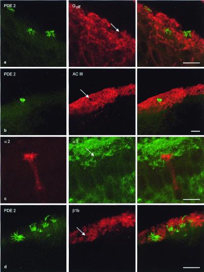
Figure 5.
Components of the olfactory cAMP-signaling pathway are absent from α2-positive neurons. (a Left) Cilia of three PDE2-positive cells; plane of section is slightly oblique. (Middle) Golf/Gs-positive cilia form a continuous layer; arrow indicates the position of one PDE2-positive ciliary bundle in the red “lawn.” (Right) Merged image, no colocalization is observed. (b Left) Cilia of a PDE2-positive cell; (Middle) ACIII-positive cilia form a continuous layer with a hole (arrow); (Right) merged image; no colocalization. (c Left) Anti-α2 immunofluorescence. (Middle) α3-positive cilia; note the hole (arrow). (Right) Merged image, no colocalization. (d Left) PDE2-positive cilia. (Middle) β1 is found throughout the ciliary layer; arrow indicates the position of a PDE2-positive ciliary bundle. (Right) Merged image, no colocalization. Scale bars = 10 μm.

