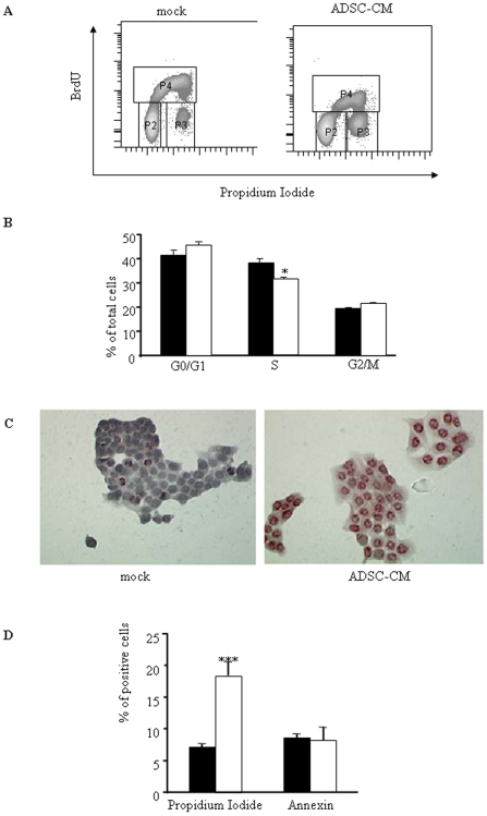
Figure 2. Human ADSCs inhibit cell proliferation and induce cell death in pancreatic cancer cells.
A. Capan-1 cells were cultured with or without ADSCs conditioned medium was added. 48 hours later, proliferation and DNA content were analyzed by flow cytometry using BrdU incorporation and propidium iodide as described in Material and Methods. Dot plots are representative of 3 independent experiments performed with ADSCs sampled from different donors. B. Quantification of cell-cycle analysis using ModFit software. Capan cultured in control conditions were represented in black bars in contrast to Capan treated with ADSC-CM represented in open bars. Values are means±S.E. of three separate experiments with ADSCs from different donors. C. Capan-1 cells were seeded in 4-chamber slides for 48 h with or without ADSC-CM. DNA fragmentation was measured by TUNEL (representative of three separate experiments). D. Capan-1 cultured in control medium (black bars) or treated with ADSC supernatants (open bars) were assayed for Annexin and propidium iodide labelling as described in Material and Methods. Values are means±S.E. of three separate experiments with ADSCs from different donors. ***: p<0.001.

