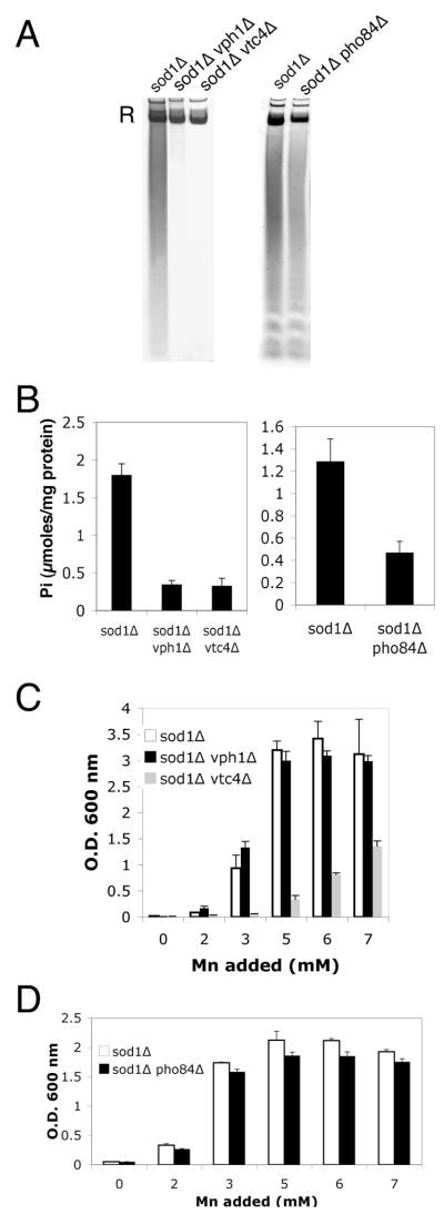
Fig. 5.
Phosphate deficient mutants of S. cerevisiae and manganese suppression of oxidative damage.
A–B) The indicated cultures were grown to early stationary phase in minimal medium containing methionine and lysine and supplemented with 3 mM MnCl2 (manganese levels that typically suppress sod1 Δ deficiency). Cell lysates were prepared for analysis of polyphosphate (A) by polyacrylamide gel electrophoresis and toluidene blue staining, or for analysis of ortho phosphate (B) by molybdate reactivity as described in Materials and Methods. “R” = RNA staining by toluidene blue. Values of orthophosphate (Pi) are the averages of three independent cultures with error bars representing standard deviation. C,D) Suppression of the sod1 Δ lysine biosynthetic defect by mM manganese was examined in triplicate cultures of the indicated strains as described in Fig. 2A. Strains used: sod1 Δ, LJ284; sod1 Δ pho84 Δ, RS001; sod1 Δ vph1 Δ, MC130; sod1 Δ vtc4 Δ, LJ286. Standard SC medium containing ≈7 mM phosphate was used for A, B left panels and for C. A,B right panels and D employed a minimal medium containing 1 mM phosphate to maximize effects of the pho84 mutation (see Materials and Methods).

