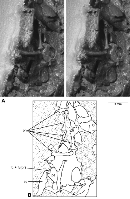
Fig. 1.
Left petrosal bone of Henkelotherium guimarotae(Gui Mam 138/76b), housed in the smaller slab (counterpart) of the type specimen. (A) Stereophotographs of the petrosal bone preserved among fragmentary skull elements and some manual phalanges. Most of the petrosal is in the resin embedding materials, not visible to the naked eye but can be visualized by computed tomography scanning. Stipple pattern indicates the coal matrix or embedding resin. (B) Explanatory drawing of (A). fc + fv(br), broken area of fenestra cochleae and fenestra vestibuli (possibly including the fossa for the stapedius muscle); pe, petrosal bone; ph, manual phalanges; sq, squamosal.

