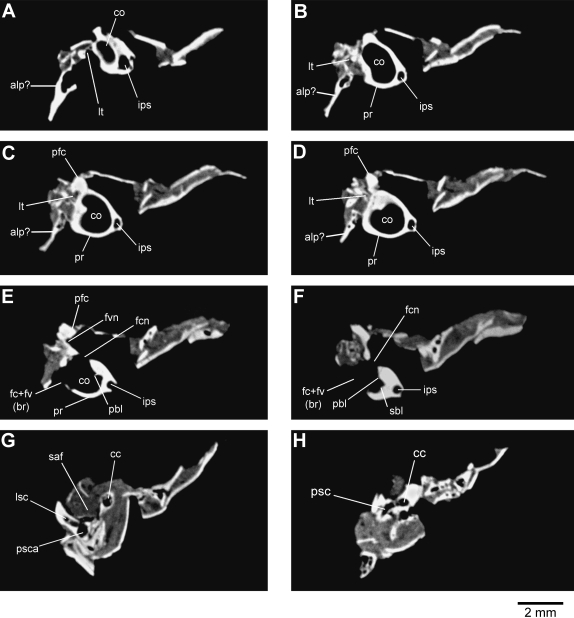
Fig. 2.
Representative computed tomography images of the left petrosal bone of Henkelotherium guimarotae (Gui Mam 138/76b). Series is from the anterior (apex of cochlea) to the posterior (posterior semicircular canal). (A) Image 054; (B) image 069; (C) image 078; (D) image 083; (E) image 096; (F) image 110; (G) image 169; (H) image 195. alp?, possible remnant of anterior lamina of petrosal; cc, crus commune; co, cochlear canal; fc + fv(br), broken area of fenestra cochleae and fenestra vestibuli (possibly including the fossa for the stapedius muscle); fcn, foramen for cochlear nerve; fvn, foramen for vestibular nerve; ips, inferior petrosal sinus; lsc, lateral semicircular canal; lt, lateral trough (incomplete); pbl, primary bony lamina; pfc, prefacial commissure; pr, promontorium; psc, posterior semicircular canal; psca, posterior semicircular canal ampulla; sbl, secondary bony lamina for basilar membrane; saf, subarcuate fossa (incomplete).

