Fig. 5.
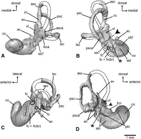
Virtual endocast of the inner ear bony labyrinth of Henkelotherium guimarotae type specimen (Gui Mam 138/76b), left side. Asterisks indicate the separation between the saccular space and the base of cochlear canal. Dark triangles indicate the separation between the utricle and saccule. (A) Anterior (rostral) view; (B) posterior view; (C) ventral view; (D) medial (endocranial) view. ac, aqueductus cochleae (endocast); asc, anterior semicircular canal (incomplete, broken segment indicated by dotted lines); asca, anterior semicircular canal ampulla; av, aqueductus vestibuli (endocast); cc, crus commune; co, cochlear duct (endocast); fc + fv(br), dashed outline indicating the broken area of fenestra cochleae, fenestra vestibuli and possibly the fossa for the stapedius muscle; fc, hypothetical position of fenestra cochleae (not preserved); fcn, foramen for cochlear nerve; fv, hypothetical position of fenestra vestibuli (not preserved); lsc, lateral semicircular canal (incomplete, broken segment indicated by dotted lines); lsca, lateral semicircular canal ampulla; pbl, basal portion of primary bony lamina for basilar membrane; psc, posterior semicircular canal; psca, posterior semicircular canal ampulla; sa?, possible saccular part of vestibule; sbl, secondary bony lamina for basilar membrane; ul, utricle.

