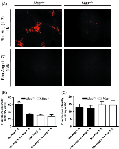
Fig. 1.
In-vitro fluorescent-labelled angiotensin-(1–7) binding in Mas+/+ and Mas−/– mouse testis. (A) Representative photomicrographs showing total binding (TB) of rhodamine-labelled angiotensin-(1–7) [Rho-Ang-(1–7)] and non-specific binding (NSB) of Rho-Ang-(1–7) in Mas+/+ and Mas−/– mouse testis. (B) Quantitative image analysis of Rho-Ang-(1–7) binding in Mas+/+ and Mas−/– mice in the intertubular compartment. (C) Quantitative image analysis of Rho-Ang-(1–7) binding in Mas+/+ and Mas−/– mice in the tubular compartment (**P < 0.0001, unpaired Student's t-test).

