Fig. 2.
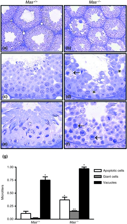
Cross-sections of seminiferous tubules in Mas+/+ (a, c and e) and Mas−/– (b, d and f) mice. Spermatogenesis is disturbed in the Mas−/– mice (b), leading to the presence of giant cells (arrow, d), vacuoles (*, d) and a large number of apoptotic cells during meiotic divisions (arrows, f). (g) Quantification of cellular volume for different cell types in Mas+/+ and Mas−/– mice. **P < 0.0001; *P < 0.01 Mas−/– vs. Mas+/+.

