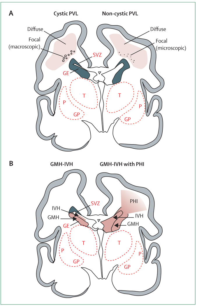
Figure 1. Cystic and non-cystic periventricular leukomalacia (PVL) and germinal matrix haemorrhage–intraventricular haemorrhage (GMH-IVH) and GMH-IVH with periventricular haemorrhagic infarction (PHI).
Coronal sections from the brain of a 28-week-old premature infant. The dorsal cerebral subventricular zone (SVZ), the ventral germinative epithelium of the ganglionic eminence (GE), thalamus (T), and putamen (P)/globus pallidus (GP) are shown. (A) The focal necrotic lesions in cystic PVL (small circles) are macroscopic in size and evolve to cysts. The focal necrotic lesions in non-cystic PVL (black dots) are microscopic in size and evolve to glial scars. The diffuse component of both cystic and non-cystic PVL (pink) is characterised by the cellular changes, as described in the text. (B) Haemorrhage (red) into the GE results in GMH, which could burst through the ependyma to cause an IVH (left). When the GHM-IVH is large, PHI might result (right).

