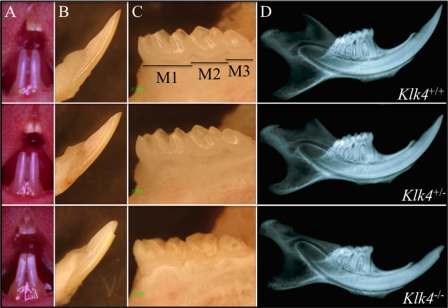
FIGURE 2.
Photographic and radiographic examination at 7 weeks. Column A, oral photographs showing incisors. Column B, lateral view of mandibular incisors following soft tissue removal. Column C, lingual view of mandibular molars (M1–M3) following soft tissue removal. Column D, radiograph of hemimandible. No differences were detected between the wild-type (Klk4+/+, top row) and heterozygous (Klk4+/−, middle row) mice at this scale. The dental enamel of the null (Klk4−/−, bottom row) mice, however, is markedly different. It is chalky in color, and the crowns have chipped off in areas of occlusal contact.

