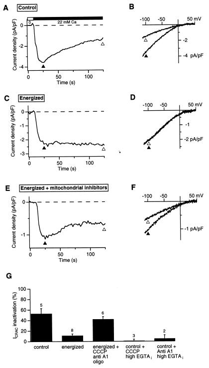
Figure 2.
Energized mitochondria prevent Ca2+-dependent inactivation of CRAC channels. ICRAC was measured in whole-cell recordings with 1.2 mM EGTA in the recording pipette. Stimulus protocol was identical to that used in Fig. 1. (A) ICRAC measured at −100 mV before and after addition of 22 mM Ca2+ using the standard whole-cell internal solution (see Materials and Methods). (B) Leak-corrected ramp currents collected at the times indicated by the triangles in A. (C, D) ICRAC recorded as in A and B with the addition of 2.5 mM malic acid/2.5 mM Na pyruvate/1 mM NaH2PO4/5 mM MgATP/0.5 mM Tris⋅GTP to the recording pipette to support mitochondrial function (“energized” conditions). (E, F) ICRAC recorded as in C and D with addition of 1 μM CCCP/2 μM antimycin A1/1 μM oligomycin to the bath. (G) CRAC channel inactivation under various conditions measured as described in Fig. 1. Inactivation under energized conditions or in the presence of 12 mM internal EGTA (last two bars) was significantly less than under the other conditions shown (unpaired Student's t test, P < 0.007). The number of cells for each condition is indicated.

