FIGURE 5.
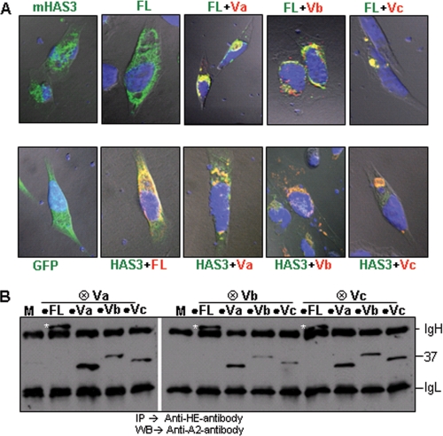
HAS1-Vs influence the localization of HAS1-FL and mHAS3. A, HAS1-Vs dominantly relocate HAS1-FL (top). As indicated in these confocal images, HeLa cells were transfected with GFP-mHAS3 or HAS1-FL alone or in combination with differentially tagged HAS1-Va, HAS1-Vb, and HAS1-Vc and stained. Green, HAS1-FL; red, all HAS1-Vs; blue, nucleus. Areas of coexpression appear as yellow. HAS1-Vs also dominantly relocate mHAS3 (lower panel). GFP alone and GFP-mHAS3 in combination with tagged HAS1-FL, HAS1-Va, HAS1-Vb, and HAS1-Vc were expressed and stained in red. B, biochemical association of HAS1-FL with each of the HAS1-Vs and among HAS1-Vs HE-tagged (⊗) HAS1-Vs co-transfected with A2-tagged (●) HAS1-FL or HAS1-Vs and mock transfection (M), as indicated in the panel. The samples were immunoprecipitated (IP) with anti-HE monoclonal antibody and immunoblotted (WB) with anti-A2 monoclonal antibody. The white asterisks represent the full-length protein on the blot. On the right of the panel, IgH and IgL represent heavy and light chain, respectively; the position of the 37-kDa size standard is indicated.

