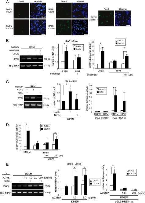
FIGURE 7.
Calcium-dependent molecular mechanism leading to IPAS gene activation. A, the intracellular calcium concentration increased 5 min after CoCl2 treatment in PRMI-cultured PC12 cells. Calcium signaling was monitored using the cells loaded with Fluo-8 AM. The data shown are representative of three experiments. The cells with A23187 treatment for 25 min were used as positive controls. Some cells with A23187 treatment underwent apoptotic DNA fragmentation. B, mibefradil (10 μm) inhibited the CoCl2-induced expression of IPAS mRNA (left and middle panels) and activated HRE-dependent luciferase activity (right panel) in RPMI-cultured PC12 cells. C, the treatment of 300 μm NiCl2 did not induce IPAS mRNA (left and middle panels) and partially enhanced HRE-dependent luciferase activity in RPMI (right panel). D, MK-801 weakly activated the reporter activity in dose dependent manner. E, A23187 induced the expression of IPAS mRNA in response to CoCl2 treatment (left and middle panels) and inhibited HRE-driven reporter activity (right panels) in DMEM. *, p < 0.05 for indicated comparison. **, p < 0.01 for indicated comparison. The data shown in the bar graphs are the averages ± S.D. of three independent experiments.

