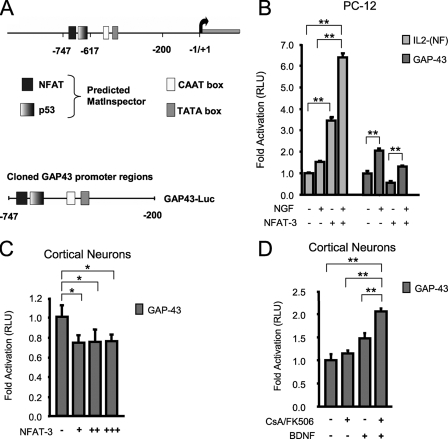
FIGURE 1.
NFAT-3 represses GAP-43 expression in PC-12 and cortical neurons. A, a depiction of the GAP-43 promoter region with the putative NFAT binding site. B, NFAT-3 represses GAP-43 transcription in PC-12 cells. Transient transfection on PC-12 cells with control IL-2-Luc, GAP-43-Luc, and EGFP-NFAT-3 expression plasmid. Cells were allowed to express EGFP-NFAT-3 for 24 h and then stimulated with 100 ng/ml NGF for an additional 24 h (see “Experimental Procedures” for DNA amounts used). RLU, relative light units. C, NFAT-3 represses GAP-43 transcription in neurons. Transient transfection was performed on cultured E16 rat cortical neurons with GAP-43-Luc reporter and increasing EGFP-NFAT-3 expression plasmid. (+, 125 ng; ++, 250 ng; +++, 350 ng) D, inhibition of the calcineurin/NFAT pathway further increases GAP-43 transcription in response to neurotrophin. Cultured E16 rat cortical neurons were transfected with GAP-43-Luc and treated with or without 10 ng/ml BDNF, 1 μm CsA, and 250 ng/ml FK506 for 16 h. All assays were performed in triplicate. The relative light unit was calculated as the ratio of firefly luciferase/Renilla luciferase signal. Signals were then normalized to reporter only transfection. Student's t test was performed to compare significant difference to reporter only transfections. p value: *, < 0.05, **, < 0.01. Error bars represent S.E.

