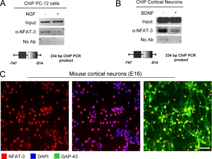
FIGURE 2.
NFAT-3 is nuclear and occupies the GAP-43 promoter in PC-12 cells, cultured neurons, and mouse brain. A, NFAT-3 occupies the GAP-43 promoter in PC-12 cells. ChIP on PC-12 cells for endogenous NFAT-3 was performed using α-NFAT-3 antibody. Cells were plated and stimulated with 100 ng/ml NGF or control for 24 h and then cross-linked and harvested. The amount of DNA recovered was measured by semiquantitative PCR. GAP-43-specific primers spanning the putative NFAT site were used. The resulting PCR amplicon containing the NFAT site of the GAP-43 promoter is depicted. No Ab, no antibody. B, NFAT-3 occupies the GAP-43 promoter in cultured cortical neurons. ChIP was performed for NFAT-3 on cultured E16 rat cortical neurons, which were treated with 10 ng/ml BDNF 1 h after plating for 5 days and harvested, and recovered DNA was measured by semiquantitative PCR. The resulting PCR amplicon containing the NFAT site of the GAP-43 promoter is depicted. C, immunocytochemistry of cultured cortical neurons shows nuclear localization for NFAT-3. Cultured E16 cortical neurons were fixed with 4% formaldehyde and probed with α-NFAT-3, α-GAP-43 antibodies, and DAPI nuclear staining. Scale bar: 20 μm.

