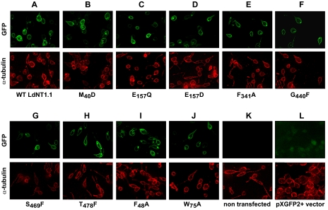
FIGURE 3.
Subcellular localization of LdNT1. 1 permeases containing crippling mutations. Each mutant was expressed as a GFP fusion at the NH2 terminus of the permease. Separate images are shown for GFP fluorescence (top row) and α-tubulin immunofluorescence (bottom row). Wild type LdNT1.1 permease was used as a positive control. Δldnt1 parasites either not transfected (K) or transfected with the pXG-GFP+2′ vector that expresses unmodified GFP (L) were also used as controls.

