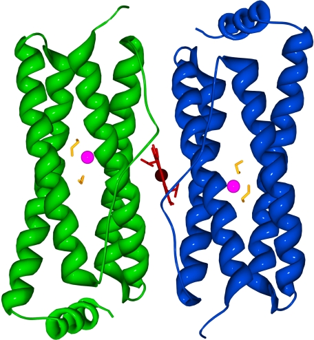
FIGURE 1.
Ribbon diagram of the structure determined for the E128R/E135R variant of E. coli BFR. The ethylene glycol molecules, zinc, and heme are colored yellow, magenta, and red, respectively. The heme group is located between the two subunits. The model is viewed from what would be the outer surface of the 24-mer.

