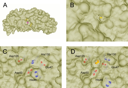
FIGURE 3.
Molecular surfaces showing the ferroxidase pore openings at the inner and outer surface. A, side view showing the relative positions of the ethylene glycol molecules and metal ion inside the protein. B, outer surface of BFR subunit dimer showing ethylene glycol molecule colored yellow in outer pore. C, inner surface of wild-type BFR showing previously observed conformations of His46, Glu47, and His130 in the closed state. D, inner surface of BFR subunit dimer showing ethylene glycol molecule in inner pore and alternate conformations of His46, Glu47, and His130 in the open state.

