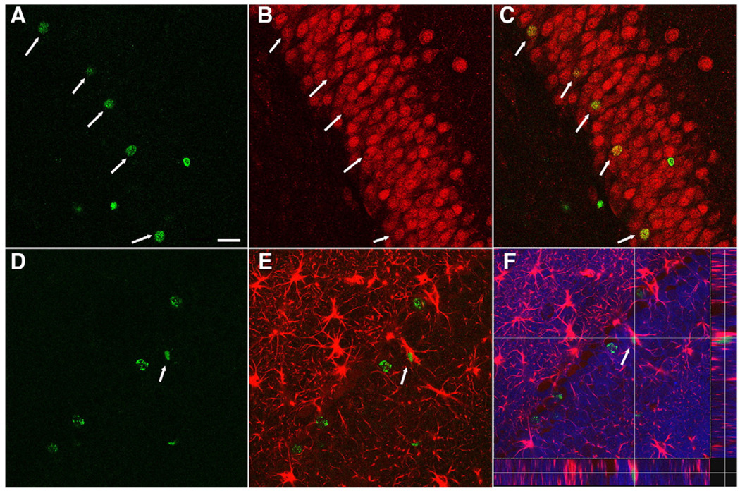
Fig. 4.
Newly generated cells in the DG following injury differentiate into both neurons and astrocytes. Confocal microscopic images of the DG at 4 weeks following injury showing double-labeling of BrdU+ cells with the neuronal marker NeuN and the astrocytic marker GFAP. (A–C) Arrows indicate BrdU-positive cells (green, A) in the DG were co-stained with the mature neuronal marker NeuN (red, B) and merged as yellow (C). (D–F) Arrow indicates co-localization of a BrdU-labeled cell (green, D) with GFAP (red, E) in the granular cells layer throughout z-axis (F, DAPI-blue). Scale bar=50 µm.

