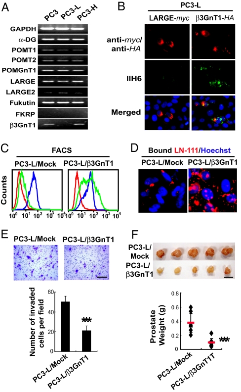
Fig. 2.
Expression of β3GnT1 directs the formation of the key laminin-binding glycans and suppresses tumor formation. (A) RT-PCR analysis of various transcripts encoding proteins implicated in the synthesis of α-DG and its functional glycosylation. (B) β3GnT1 but not LARGE expression restores laminin-binding glycans in PC3-L cells, as detected by IIH6. (C) Analysis of the expression of laminin-binding glycans (VIA4-1 in green) and α-DG (6C1 in blue) in PC3-L cells stably expressing β3GnT1 (PC3-L/β3GnT1) and mock-transfected PC3-L cells. Red is control. (D) Immunofluorescent detection of soluble laminin-111 binding to the stable transfectants. (E) Invasion assay of PC3-L/β3GnT1 and PC3-L/Mock using a transwell. The invaded cells were stained with crystal violet and counted. Small dots are pores on the membrane. (Scale bar: 200 μm.) (F) Primary tumors formed by PC3-L/Mock and PC3-L/β3GnT1 in orthotopic SCID mouse prostates 4 weeks after inoculation of the cells. (Scale bar: 1 cm.)

