Figure 4.
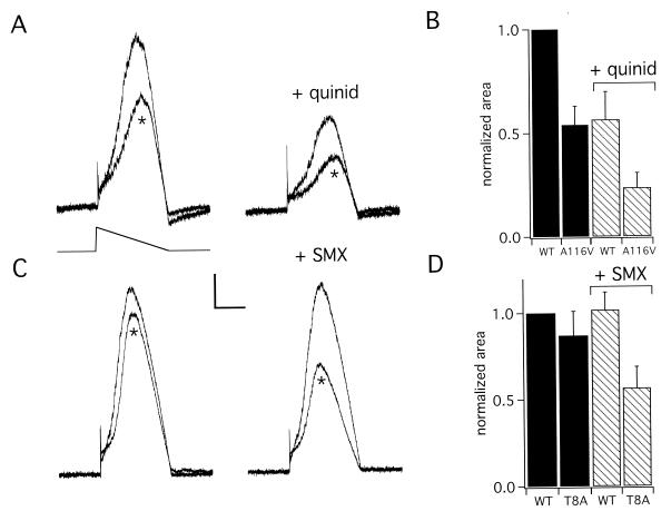
Missense mutations and drug inhibition combine to diminish potassium flux. Simulated cardiac action potentials were used to study channels formed with wild-type MiRP1, A116V-MiRP1 and T8A-MiRP1 with HERG in CHO cells. (A) Whole-cell currents for simulated action potentials with wild-type MiRP1 and A116V-MiRP1 (*) in the absence and presence of 0.5 μg/ml quinidine (+quinid). Scale bars represent 1.3 pA/pF and 0.8 s. (Inset) Protocol: holding −80 mV, voltage ramp from 40 to −80 mV, dV/dt = −71 mV/s. (B) Normalized potassium flux assessed by measurement of area under the curve for simulated action potentials in cells expressing wild-type MiRP1 (WT) or A116V-MiRP1 (A116V) in the presence of 0.5 μg/ml quinidine (+quinid) normalized to wild-type channels without drug. Currents were divided by cell capacitance before normalization. Each bar represents the mean ± SEM for 6–10 cells. (C) Whole-cell currents for simulated action potentials with wild-type MiRP1 and T8A-MiRP1 (*) in the absence and presence of 300 μg/ml SMX. Scale bars represent 1.6 pA/pF and 0.8 s. (D) Normalized potassium flux assessed by measurement of area under the curve for simulated action potentials in cells expressing wild-type MiRP1(WT) and T8A-MiRP1 (T8A) in the presence of 300 μg/ml SMX normalized to wild-type channels without drug. Currents were divided by cell capacitance before normalization. Each bar represents the mean ± SEM for 7–10 cells.

