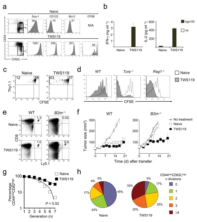
Figure 3. Wnt signaling promotes the generation of TSCM.
CFSE-labeled, naive pmel-1 CD8+ T cells were primed in vitro with CD8+ T cell-depleted splenocytes pulsed with 1 μM hgp10025–33, in conjunction with 10 ng ml−1 IL-2 and 7 μM TWS119. a, Flow cytometry analysis of TWS119-treated and naive pmel-1 cells four days following T cell activation. b, Cytokine release assay of sorted CD44lowCD62Lhigh TWS119-treated and naive pmel-1 cells five days after antigenic stimulation. Data are represented as mean +/− SEM. c and d, CFSE dilution of sorted CD44lowCD62Lhigh TWS119-treated and naive thy 1.1+ (c) or ly5.1+ (d) pmel-1 cells one month after transfer into WT (c) and sublethally-irradiated WT, or Tcra−/−, or Rag1−/− mice (d). Data are shown on thy 1.1+ (c) or ly5.1+ (d) CD8+ lymphocytes. e, Flow cytometry analysis of sorted TWS119-treated and naive ly5.1+ pmel-1 cells one month after transfer into WT or B2m−/− mice. Data are shown on ly5.1+, CD8+ lymphocytes. f, Tumor treatment of myeloablated WT or B2m−/− mice bearing B16 tumors established for 7 days. Mice received age-matched, lineage-depleted bone marrow cells. On the following day, 106 CD44lowCD62Lhigh TWS119-treated or naive pmel-1 cells were transferred in conjunction with exogenous IL-2.g and h, Flow cytometry analysis of CFSE-labeled, sorted CD44lowCD62Lhigh TWS119-treated and naive ly5.1+ pmel-1 cells one month after transfer into sublethally-irradiated WT mice. Data are represented as the percentage of CD44lowCD62Lhigh cells as a function of CFSE dilution of two independent experiments (g) and as fraction of cells with any given number of divisions (h). All data are representative of at least two independently performed experiments.

