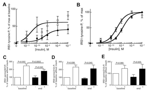
Figure 1.
Phosphorylation of IRS1 at tyrosine (A,B) at serine-307 (C) and serine-312 (D,E). (A,B) Adipocytes obtained at baseline (open circles) and at the end (closed circles) of the high-calorie overeating period (A) or from patients with type 2 diabetes (closed circles), age 35–74 years, average 57 years; BMI 28–43 kg/m2, average 35 kg/m2 (n = 15) compared with nondiabetic controls (open circles), age 31–82 years, average 55 years; BMI 23-33 kg/m2, average 26 kg/m2 (n = 16) (B), were incubated with the indicated concentration of insulin for 10 min, when incubations were terminated and equal volumes of whole cells were subjected to SDS-PAGE and immunoblotting for phosphotyrosine. The phosphorylation of IRS1 is expressed as a percentage of maximal insulin effect at the start (baseline) of the high-calorie diet, mean ± SE (n = 6) (A), or as a percentage of maximal insulin effect in control cells and in diabetic cells, respectively, mean ± SE (B). Samples from the same subject were analyzed on the same gel. The curves were statistically different: P = 0.0009 (A); P < 0.0001 (B), F-test. (C–E) Adipocytes obtained at baseline (open bars) and at the end (closed bars) of the high-calorie overeating period were incubated with (+) or without (−) 100 nmol/L insulin, as indicated. After 10 min (C,E) or 30 min (D) incubations were terminated and equal volumes of whole cells were subjected to SDS-PAGE and immunoblotting for phosphorylation of IRS1 at serine-307 (C) or serine-312 (D,E). Whereas insulin-stimulated phosphorylation of IRS1 at serine-307 is maximal after 10 min, the phosphorylation at serine-312 requires 30 min (10); we therefore incubated for 10 or 30 min, as indicated. The phosphorylation of IRS1 in response to insulin is expressed as a percentage of baseline phosphorylation without insulin; mean ± SE (C, n = 6; D, n = 3; E, n = 6). Samples from the same subject were analyzed on the same gel. Paired Student t test was used for statistical analysis.

