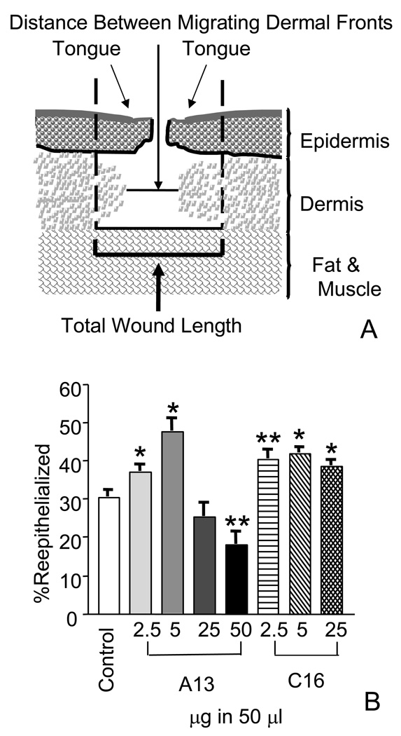
Figure 1.
Laminin-derived peptides increased reepithelialization when applied topically. A) Diagram of a full thickness punch wound. The reepithelialization as measured by % coverage and contraction as measured by % granulation were used to calculate wound closure. % reepithelialized= Tongue + Tongue /Total Wound Length ×100. % granulated = 1- Distance Between Migrating Dermal Fronts/Total Wound Length ×100 B) Quantitation of wound reepithelialization in wounds harvested at day four. Wounds were treated with peptides ranging from 2.5 to 50 µg in 50 µl. Treatment with A13 at the 2.5 and 5 µg in 50 µl level, wounds showed a significant increase in the increase in epithelial coverage of the wound, 7% (±2.5) and 17% (±3.8), respectively. At the higher dose of 50 µg in 50 µl, A13 showed a statistically significant level of inhibition of reepitheilization at 12% (±3.8). C16 peptide treatment also increased reepithelialization in topical treatments. At all doses, a significant increase in the levels of reepithelialization was observed. At 2.5 µg, a 10% (±2.9) increase in coverage was observed. At 5 µg, coverage increased to 11% (±2.4) and at higher doses, 25 µg in 50 µl, an 8% (± 2.2) increase in coverage was observed. Measurements are expressed as the mean % increase ± SEM. (*p≤0.008, **p≤0.02).

