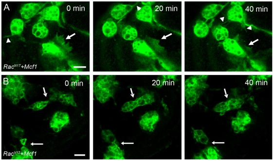
Figure 6. Embryos with defective Rac activity evade Mcf1 mediated paralysis.
(A) Stills from a movie (see Video S9) of hemocytes expressing RacN17 following injection of Mcf1. Hemocytes expressing RacN17 are localised at the anterior of the embryo and have decreased lamellipodia formation and movement compared to wild-type cells. However, despite these defects RacN17 hemocytes fail to freeze after Mcf1 injection and continue to form small dynamic membrane ruffles (arrow) and filopodia (arrowheads). (B) Time-lapse movie stills (see Video S10) showing constitutively active RacV12 expressing hemocytes following Mcf1 injection. RacV12 expression in hemocytes causes reduced lamellipodia formation and migration when compared to wildtype cells. When exposed to Mcf1 these cells fail to display the freezing phenotype and like the RacN17 expressing cells continue to make small dynamic protrusions (arrows). Scale bars represent 10 µm. Elapsed time is indicated in the upper right corner.

