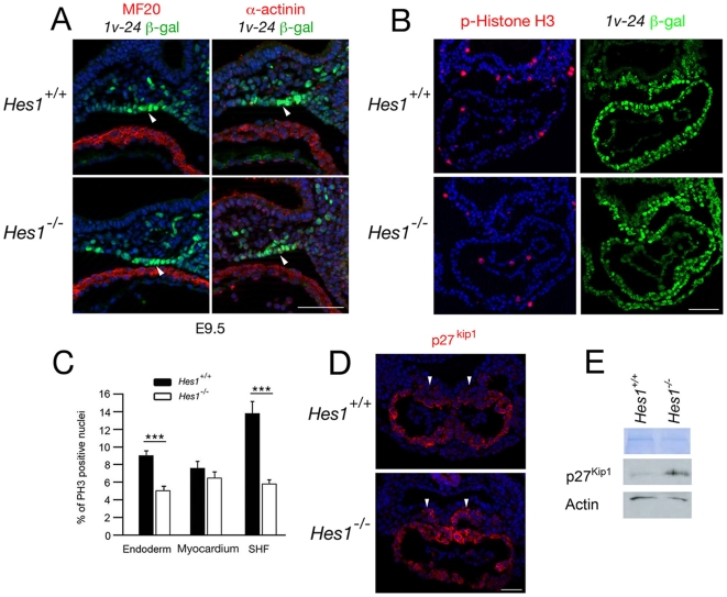
Figure 7. Impact of loss of Hes1 on differentiation and proliferation in the second heart field.
(A) Immunochemistry with anti-MHC (left), anti-α-actinin (right) and anti-β-galactosidase antibodies in transverse sections through the caudal pharyngeal region of an E9.5 embryo carrying the Mlc1v-nlacZ-24 transgene. Note the β-galactosidase positive α-actinin and MF20 negative cells in the dorsal pericardial wall (arrowheads). Nuclei are labeled with Hoechst (blue). (B) Immunochemistry with anti-phospho-Histone H3 and anti-β-galactosidase antibodies in paraffin sections of E8.5 Hes1 +/+ and Hes1 −/− embryos carrying the Mlc1v-nlacZ-24 transgene. (C) Histogram comparing the percentage of phospho-Histone H3 positive nuclei in pharyngeal endoderm, myocardium and SHF of Hes1 +/+ and Hes1 −/− embryos. A decrease in phospho-Histone H3 positive nuclei is observed in Hes1 −/− SHF and endoderm (p<0.001, Student's t-test). (D) Immunochemistry with an anti-p27kip1 antibody in paraffin sections at E8.5. Note that p27kip1 is expanded in the SHF of Hes1 −/− hearts (arrowheads). Nuclei are labeled with Hoechst (blue). (E) Western blot of microdissected heart and ventral pharyngeal regions of Hes1 +/+ and Hes1 −/− embryos showing elevated p27kip1 protein levels in the absence of Hes1. Scale bars (A): 50 µm; (B): 100 µm; (D) 50 µm.

