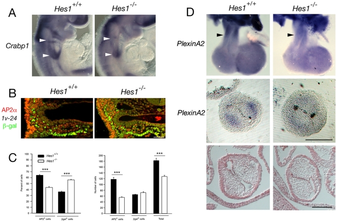
Figure 8. Loss of Hes1 impairs neural crest development.
(A) In situ hybridization at E9.5 showing decreased Crabp1 transcript accumulation in the caudal pharyngeal region of Hes1 −/− compared to Hes1 +/+ embryos (arrowheads). (B) Immunochemistry with anti-AP-2α (red) and anti-β-galactosidase (green) antibodies in a transverse section through the caudal pharynx of an E9.5 embryo carrying the Mlc1v-nlacZ-24 transgene. (C) Histograms showing the percentage of β-galactosidase positive nuclei and the percentage of AP-2α positive nuclei in the pharyngeal region of Hes1 +/+ and Hes1 −/− embryos (left) and the numbers of total, β-galactosidase positive and AP-2α positive nuclei (right). Note the decrease in the number of AP-2α positive cells in Hes1 −/− embryos (p<0.001, Student's t-test). (D) In situ hybridization at E11.5 showing decreased PlexinA2 transcript accumulation in the OFT of Hes1 −/− compared to Hes1 +/+ hearts (black arrowheads) in wholemount (top) and transverse sections (middle). Histological analysis reveals normal OFT cushion morphology in Hes1 −/− embryos at E11.5 (bottom). Scale bars (B): 50 µm; (D): 200 µm.

