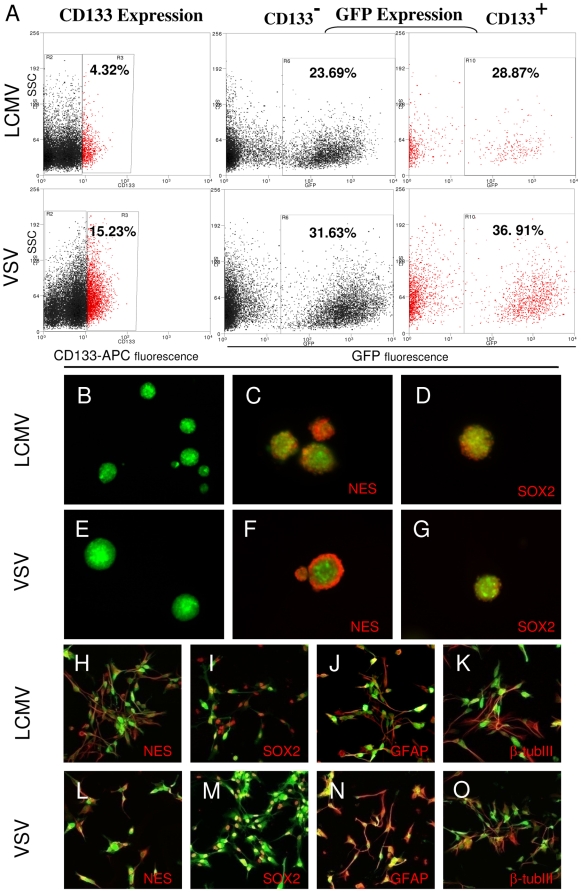
Figure 5. Lentiviral vectors transduce cancer stem-like cells.
Intracranial gliomas were infected with LCMV-GP or VSV-G pseudotyped lentiviral vectors expressing eGFP 3–4 weeks after tumor implantation. Tumors were excised when large lesions appeared on MRI and were enzymatically dissociated. The transduction of CD133 positive cells was measured by flow cytometry. (A) LCMV-GP and VSV-G pseudotyped vectors transduce CD133 positive and negative tumor cells. The fraction of transduced (GFP-positive) cells is slightly higher in CD133 positive cells (right column) compared to CD133 negative cells (middle column). GFP+ cells were sorted, cultured in the presence or absence of serum and analyzed by fluorescence (B-G) or confocal microscopy (H-O). LCMV-GP (B) and VSV-G (E) transduced cells form spheroids upon culture in serum-free neural basal medium supplemented with EGF and bFGF. Transduced spheroids express the neural stem cell markers nestin (C,F) and SOX2 (D,G). Transduced cells cultured in serum-containing medium express the stem cell markers nestin (H,L) and SOX2 (I,M), but also the differentiation markers GFAP (J,N) and beta-tubulinIII (K,O). The pictures C,D,E,F,H-O show overlay of the virus-delivered transgene (eGFP, green) and detected antigen (Alexa-647, red).

