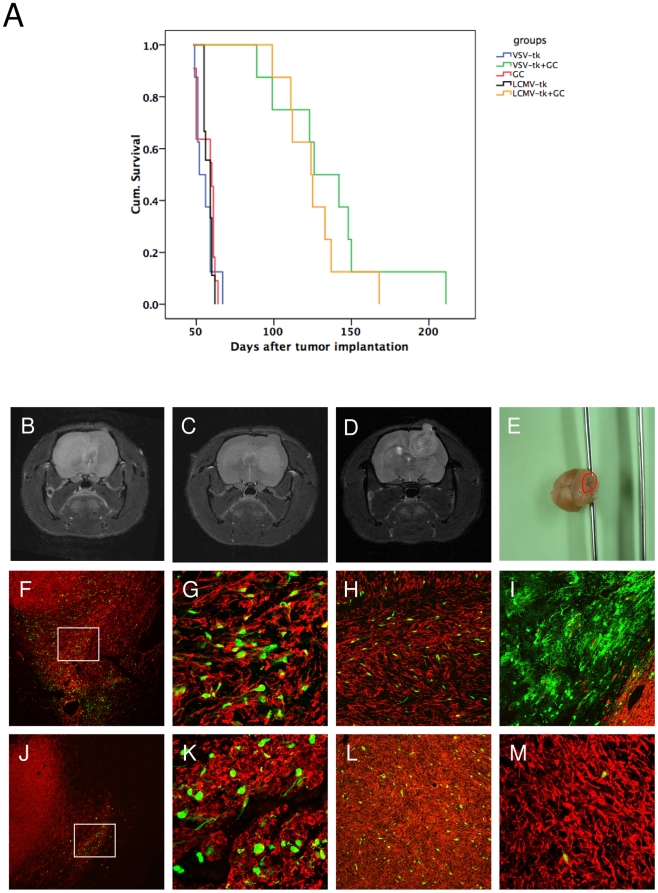
Figure 7. Therapeutic efficiency of LCMV-GP and VSV-G pseudotyped lentiviral vectors in vivo.
Intracranial gliomas were injected with LCMV-GP or VSV-G pseudotyped lentiviral vectors expressing HSV-1-tk fused to eGFP 3 weeks after tumor implantation. 7 days after vector infection, animals in both treated groups and in one control group were treated with ganciclovir for 30 days. (A) Kaplan-Meier survival curve. The survival benefit for both treatment groups compared to control groups was statistically significant (P<0.001; log-rank test). There was no significant difference in survival between the two treatment groups. (B-D) Representative MRI (T2 RARE) of recurrent tumors in the LCMV- (B,C) and VSV-treated (D,E) group. (B) Invasive contralateral recurrence. (C) Invasive local and contralateral recurrence. (D) More circumscribed local recurrence. (E) Macroscopic picture of a rat brain with a recurrence in the cerebellum (red circle), treated with VSV-G pseudotyped vectors and GC. (F-M) Sections of recurrent tumors were stained with antibodies against human-specific nestin and analyzed by fluorescence (F,J) or confocal microscopy (G-I,K-M). Pictures show overlay of nestin (red) and eGFP transgene (green). (F-I) Recurrent tumors of animals treated with VSV-G pseudotyped lentiviral vectors. (F) Recurrent tumor with GFP-positive cells in the invasive area. (G) Higher magnification of (F). (H) GFP-positive tumor cells in the corpus callosum region. (I) GFP-positive normal brain cells at the tumor border. (J-M) Recurrent tumors of animals treated with LCMV-GP pseudotyped lentiviral vectors. (J) GFP-positive tumor cells in residual small lesion from the primary tumor. The recurrent tumor is growing from the contralateral hemisphere over the corpus callosum to the ipsilateral hemisphere (arrows). (K) Higher magnification of (J). (L) GFP-positive tumor cells in the solid ipsilateral recurrent lesion. (M) Few GFP-positive cells in a contralateral recurrent tumor. F,J: Magnification 40×. G,K,M: Magnification 200×. H,I,L: Magnification 100×.

