
Figure 2. Shikonin dosage effects on proteasome inhibition and cell death induction in PC-3 and H22 cells.
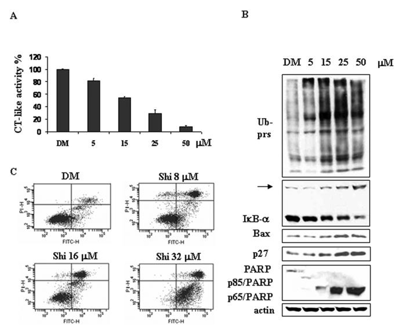
A-B, Human prostate cancer PC-3 cells were treated with either solvent DMSO (DM) or indicated concentrations of shikonin for 6 h, followed by measuring the inhibition of the protesomal CT-like activity using fluorescent substrate Suc-LLVY-AMC (A) and Western blotting analysis using specific antibodies against ubiquitinated proteins, IκB-α Bax, p27 and PARP (B). Columns, means of independent triplicate experiments; bars, SD. Molecular weight of IκB-α Bax and p27 is 37, 23 and 27 kDa, respectively. An ubiqutinated form of IºB-± (~56 kDa)30 was indicated by an arrow. Full length PARP is 116 kDa, the cleaved fragments of PARP are 85 kDa or 65 kDa. Actin was used as loading control. C, Murine hepatoma H22 cells were treated with either DMSO (DM) or 8, 16 or 32 μM shikonin for 24 h, followed by Annexin V-FITC binding assay. The lowerright part (Annexin V-FITC + / PI -) was considered as early stage of apoptotic cells and upright part (Annexin V-FITC + / PI +) was considered as late stage of apoptotic cells. The lowerleft part (Annexin-FITC -/PI -) was considered as viable cells and the upleft part (Annexin V-FITC - / PI +) was considered as necrotic cells.