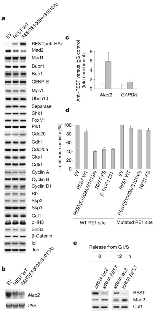
Figure 2. Mad2 is a transcriptional target of REST.
a, U-2OS cells were infected either with an empty lentivirus (EV) or with lentiviruses expressing HA-tagged wild-type REST or HA-tagged REST(E1009/S1013A). After treatment with nocodazole for 15 h, mitotic cells were harvested and analysed by immunoblotting for the indicated proteins. b, Mad2 mRNA was assessed by northern blotting in U-2OS cells treated as in a. c, Chromatin immunoprecipitation (ChIP) assay with an anti-REST antibody (filled columns) in U-2OS cells. Quantitative real-time PCR amplifications were performed with primers surrounding the RE1 site in the Mad2 promoter. The value given for the amount of PCR product present from ChIP with control IgG (open columns) was set as 1. GAPDH primers were used as a negative control. d, U-2OS cells were transfected with an empty vector (EV), HA-tagged REST proteins or a dominant-negative β-TrCP1 mutant (FLAG–β-TrCP1 DN) together with a luciferase reporter linked to a Mad2 genomic fragment containing either a wild-type (WT) or a mutated RE1 site. Prometaphase cells were collected and the relative luciferase signal was quantified. The value given for luciferase activity in EV-transfected cells was set at 100%. e, IMR-90 cells were transfected twice with siRNA molecules to a non-relevant mRNA (lacZ) or to REST mRNA and synchronized in G2 by release from an aphidicolin block for the indicated durations30. Cells were then lysed and immunoblotted. Where present, error bars represent s.d. (n = 3).

