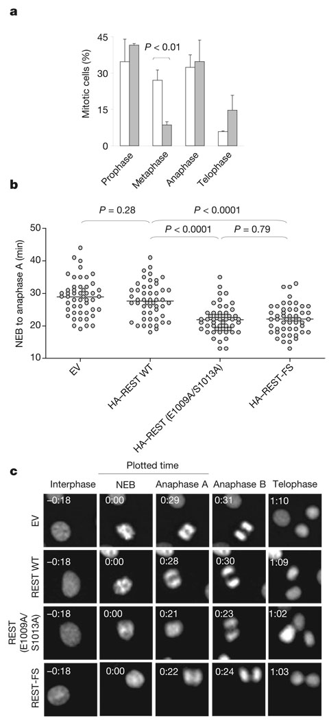
Figure 3. Failure to degrade REST causes defects in the mitotic checkpoint.
a, HCT116 cells were infected either with an empty lentivirus (EV, open columns) or with a lentivirus expressing REST(E1009/S1013A) (filled columns). At 48 h after infection, cells were fixed and stained with 4,6-diamidino-2-phenylindole and an anti-α-tubulin antibody to reveal DNA and the mitotic spindle, respectively. Error bars represent s.d. (n = 3). b, NIH 3T3 cells stably transfected with enhanced green fluorescent protein-labelled histone H2B were infected with an empty lentivirus or with lentiviruses expressing the indicated HA-tagged proteins. The average time from nuclear envelope breakdown (NEB) to anaphase onset was measured by time-lapse microscopy. Each symbol in the scatter plot represents a single cell. c, Representative fluorescence videomicroscopy series from b; numbers in the top left are times (h:min).

