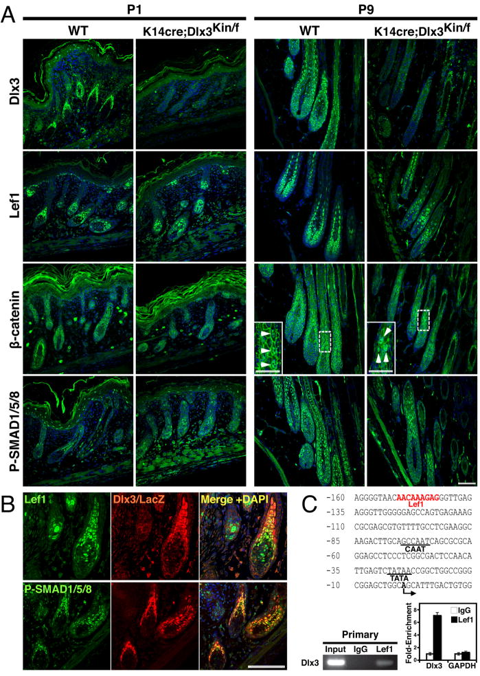
Fig. 5. Follicular signaling in the regulation of Dlx3 expression.
(A) Expression of Lef1, β catenin and SMAD1/5/8 were determined in control and conditional knockout skin to analyze effects of absence of epithelial Dlx3 at P1 and P9 anagen stages. (B) Co-localization of Dlx3/LacZ with Lef1. The skin sections of wild type (Dlx3Kin/+) at P1 were stained with by anti-Lef1, anti-phospho SMAD1/5/8, and anti-β-galactosidase antibodies (red, middle), and the merged images are shown with DAPI staining. (C) Lef1 direct binding in vivo to the Dlx3 promoter was demonstrated by ChIP assays using Lef1 antibody. Putative Lef1 binding site in the mouse Dlx3 promoter sequence (−160 to +15bp) is indicated in red letters. CCAAT box and TATA box are underlined. The transcriptional start site is indicated by an arrow. Bottom left shows gel image of ChIP assay and right panel shows the specificity of the ChIP assays, which was determined by both control IgG antibody and a set of PCR primers for GAPDH. Scale bar, 50μm.

