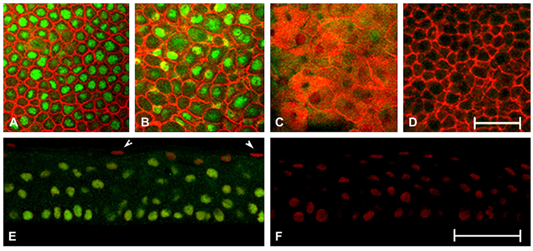
Fig. 1.
Immunostaining for ΔNp63 in normal human tissue. (A–D) Representative XY slices of whole mount human corneal epithelium double-labeled with a rabbit polyclonal antibody directed against the N terminus of ΔNp63 (green) and phalloidin (red). (A) Basal cell layer; (B) wing cell layer; (C) superficial cell layer; (D) negative control, primary antibody omitted. (E, F) 10 µm cryosections double-labeled with anti-rabbit ΔNp63 (green) and PI (red). (E) Human central corneal epithelium. Arrows indicate DNp63-negative, PI-positive cells; (F) negative control, primary antibody omitted. Whole mount experiments were repeated three independent times; cryostat sectioned experiments were repeated three independent times. Scale: 50 µm.

