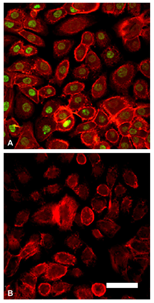
Fig. 3.
Immunostaining for ΔNp63 in hTCEpi cells. (A) hTCEpi cells grown on collagen-coated glass coverslips double-labeled with ΔNp63 (green) and rhodamine-conjugated phalloidin (red). All cells were positive for nuclear p63. (B) Negative control, primary antibody omitted, counterstained with phalloidin. Scale: 45 µm.

