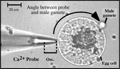
Figure 1.
Micrograph from a typical Ca2+ flux measurement during maize IVF. The Ca2+-selective vibrating probe was positioned approximately 1 μm from the surface of the egg cell at its nearest vibration point. The probe oscillated with an excursion of 10 μm away from the cell (Osc.). The angle between the probe and the adhering male gamete is indicated by the black arrow.

