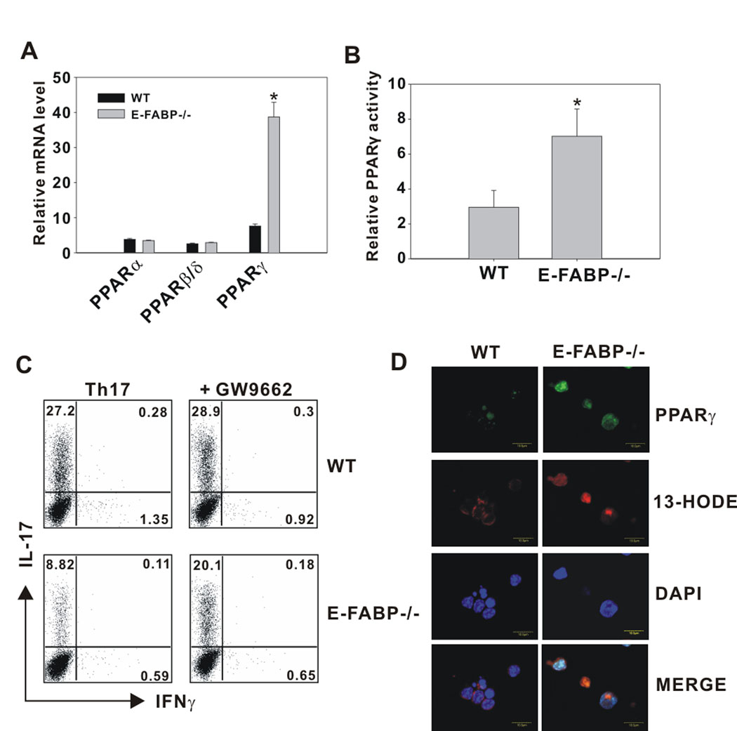
Figure 5. E-FABP deficiency is associated with elevated PPARγ expression and activity.
A, Naïve CD4+ T cells purified from WT and E-FABP-deficient mice were cultured with anti-CD3 plus anti-CD28 for 3 d. Relative expression of PPARα, PPARδ/β and PPARγ was analyzed by quantitative real-time PCR. Expression was normalized to β-actin and presented as the level of mRNA expression relative to a non-stimulated WT naïve T cell control. Data shown are representative of three similar experiments (* p< 0.05). B, In vitro cultured T cells from WT and E-FABP-deficient mice were stimulated for 3 d. PPARγ activity in nuclear lysates was measured by relative comparison with an unstimulated WT control (* p < 0.05). C, Naïve CD4+ T cells were cultured for 3 d under Th17 conditions in the presence or absence of the PPARγ antagonist GW9662 (5µM). T cells were recovered and re-stimulated with PMA plus ionomycin for 5 h and analyzed for IL-17 and IFNγ expression by intracellular labeling and flow cytometric analysis. D, In vitro cultured T cells from WT and E-FABP-deficient mice were incubated with 13-HODE-biotin and labeled with streptavidin (red, Alexa Fluor 5680) and FITC conjugated anti- PPARγ (green). Nuclei were stained with DAPI (blue).

