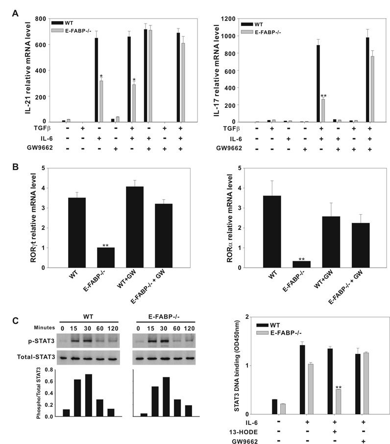
Figure 6. The reduced Th17 differentiation exhibited by E-FABP-deficient T cells is mediated by PPARγ.
A, WT and E-FABP-deficient naïve T cells were stimulated with anti-CD3 and anti-CD28 in the presence of combinations of cytokines +/− GW9662 (GW), as indicated. Data shown are representative of three similar experiments (* p < 0.05, ** p < 0.01). B, Expression of RORγt (left panel) and RORα (right panel) mRNA by naïve T cells, cultured as under Th17 conditions +/− GW9662 , was analyzed by real-time RT PCR (** p < 0.01). C, Left panel, naïve CD4+ T cells from WT and E-FABP were purified and stimulated with IL-6 at the indicated time points on plate-bound anti-CD3 and soluble anti-CD28. Cell lysates were analyzed by Western blot using Abs specific for the phosphorylated form of STAT3, or total STAT3, as indicated. The histogram represents ratio of phosphorylated STAT3 to total STAT3 band density. Right panel, CD4+ T cells from WT and E-FABP-deficient mice were treated without or with 13-HODE +/− GW9662 for 3h followed by a 20 min stimulation with IL-6. STAT3 DNA binding was assayed as described in Material and Methods. The data are presented as mean ± standard deviation of triplicate samples (** p < 0.01).

