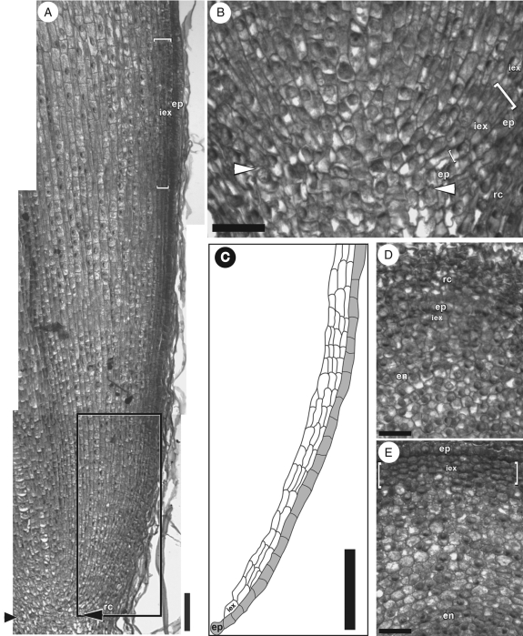
Fig. 2.
Iris germanica adventitious root tips in (A, B) longitudinal and (D, E) transverse sections. (A) Tip of a 4-mm-long adventitious root. Arrowheads indicate the distal extremity of the root apical meristem. (B) Enlargement of the apical meristem area (arrowheads as in A). The epidermis is immature and the number of developing immature exodermal cell files varies (within square brackets). (C) Tracing of the immature epidermal (grey) and exodermal (white) cells, located in the rectangle outlined in (A). Note the variable number of immature exodermal cell files in this region. (D) Section 50 µm from the tip of the root proper. Note the lack of regular radial alignments of cells across the young cortex. The root cap is thick at this distance. (E) Section 200 µm from the tip of the root proper. A boundary between the epidermis and immature exodermis is noticeable at this distance. There are two to four layers of cells in the immature exodermis, which are characterized by a lack of intercellular air spaces (within square brackets). Abbreviations: Rc, root cap; ep, epidermis; iex, immature exodermis; en, endodermis. Scale bars: (A, C) = 100 µm; (B, D, E) = 50 µm.

