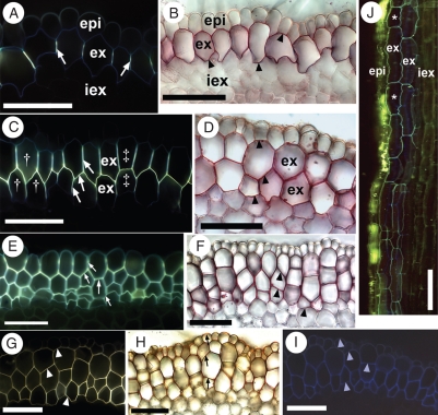
Fig. 3.
Photomicrographs of the outer part of I. germanica roots in (A–I) cross- and (J) longitudinal section. Values in millimetres refer to distances from the root tips. (A) 15 mm. Stained with berberine hemisulfate–aniline blue. Casparian bands (white arrows) fluoresced yellow and occupied the anticlinal walls of the outermost exodermal layer. (B) 15 mm. Stained with Sudan red 7B. Suberin lamellae (black arrowheads) appeared as red rings in the walls of the outermost exodermal layer. (C) 70 mm. Stained with berberine hemisulfate–aniline blue. The ccCb (white arrows) was Y-shaped (in cells labelled with †) or H-shaped (in cells labelled with ‡). (D) 70 mm. Stained with Sudan red 7B. Cells in the two mature exodermal layers contained suberin lamellae (black arrowheads). (E) 100 mm. Stained with berberine hemisulfate–aniline blue. The ccCb (white arrows) filled the anticlinal and tangential walls of cells in the multiseriate exodermis. (F) 100 mm. Stained with Sudan red 7B. Cells in the multiseriate exodermis all contained suberin lamellae (black arrowheads). (G) 100 mm. Stained with Fluorol yellow 088. Suberin (white arrowheads) fluoresced yellow in exodermal cell walls. (H) 100 mm. Stained with phloroglucinol–HCl. Lignin (black arrows) appeared reddish-orange in the walls of epidermal and exodermal cells. (I) 70 mm. Autofluorescence with UV light. Walls of the epidermis and exodermis autofluoresced faint blue (light blue arrowheads). (J) 70 mm. Epidermal cells containing berberine thiocyanate crystals (yellow) and a dimorphic, biseriate exodermis with blue, autofluorescent walls. Asterisks indicate short cells. Abbreviations: epi, epidermis; ex, mature exodermis; iex, immature exodermis. Scale bars = 100 µm.

