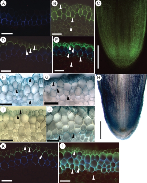
Fig. 7.
Apoplastic permeability tests on I. germanica roots with berberine or FeSO4. The photomicrographs show cross-sections of the roots unless otherwise stated. Values in millimetres refer to distances from the root tips. (A) 90 mm. Unstained, UV autofluorescent control. All exodermal walls were faint blue. (B) 60 mm. Entire section stained with berberine. Epidermal, exodermal and central cortical cell walls fluoresced yellow (white arrowheads). (C) Longitudinal section of a root tip treated externally with berberine. The walls of all cells near the tip fluoresced yellow. (D) 70 mm. The outermost Casparian bands of an intact multiseriate exodermis prevented externally applied berberine (white arrowheads) from permeating the exodermis and central cortex. (E) 70 mm. A punctured multiseriate exodermis allowed berberine to stain the walls of epidermal, immature exodermal and central cortical cells (white arrowheads). (F) 80 mm. Entire section treated with FeSO4 followed by K4[Fe(CN)6]·3H2O. Ferric ions precipitated in all epidermal and exodermal walls and appeared blue (black arrowheads). (G) 70 mm. Following an external treatment, ferric ions were detected in mature exodermal walls (black arrowheads). (H) Longitudinal section of a root tip treated externally with FeSO4 followed by K4[Fe(CN)6]·3H2O. The ferric ions entered the cortex of the tip readily. (I) 90 mm. Ferric ions, as evidenced by blue precipitates (black arrowheads), were blocked from permeating the exodermal Casparian bands. (J) 90 mm. A punctured multiseriate exodermis allowed the ferric ions limited entry to the central cortex (black arrowheads). (K) 70 mm. Intact root incubated in water and then berberine. Berberine (white arrowheads) did not permeate the Casparian bands in the first exodermal layer. (L) 100 mm. Intact root incubated in FeSO4 and then berberine. Berberine stained the walls of all mature and immature exodermal cells, and walls of cortical parenchyma (white arrowheads). Scale bars: cross-sections = 50 µm; longitudinal sections = 500 µm.

