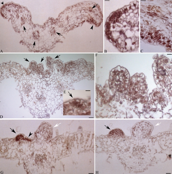
Fig. 4.
Localization of Ha-L1L transcripts in cross-sections of epiphyllous leaves (EP) of the interspecific hybrid Helianthus annuus × H. tuberosus. The presence of Ha-L1L mRNAs is evidenced as a violet-brownish staining. (A) Leaf blade proximal to the petiole junction; arrowheads indicate labelled cells grouped along the leaf margin and discontinuously distributed along the blade; arrows indicate labelled vascular bundles. (B, C) Higher magnification of (A). (D) Distal leaf; arrows indicate ectopic embryos. (E, F) Higher magnification of (D); arrow in (E) indicates incipient ectopic structure. (G, H) Distal leaf blades; arrowhead indicates a linear cluster of labeled cells; arrows indicate advanced globular embryos; white arrows indicate ectopic structures arrested in development. Scale bars: (A) = 35 µm; (B, C) = 11 µm; (D) = 45 µm; (E) = 10 µm; (F) = 16 µm; (G, H) = 35 µm.

