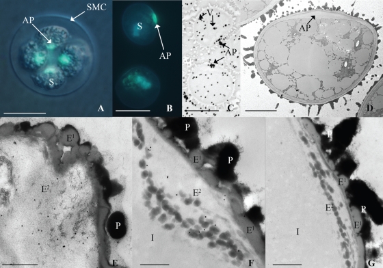
Fig. 1.
Light and electron micrographs of spores from Physcomitrella patens. (A) Spores (S) in a tetrahedral tetrad inside the spore mother cell (SMC) wall stained with aniline blue; positive reaction for callose in aperture (AP) region of spores. (B) Spores liberated from SMC with wall in early developmental stages indicated by lack of spinose morphology stained with aniline blue; positive reaction in the aperture. (C) Positive reaction to silver enhanced anti-callose immunolabel in vacuoles (V) and the aperture of mature spores. (D) TEM of unlabelled spore with expanded aperture. (E–G) TEM of spore wall at the aperture region of proximal wall consists of perine (P), outer exine (E1), inner exine (E2) and intine (I): (E) oblique tangential section through aperture at early developmental stage indicated by relatively perine-free proximal wall has positive reaction to anti-callose immunogold label in the exine; (F) longitudinal section through aperture at later developmental stage indicated by the distinct presence of intine has notable positive reaction to anti-callose immunogold label associated with dark globules in (E2); (G) longitudinal section through aperture labelled with only secondary antibody as a control. Scales bars: (A–C) = 15 µm; (D) = 5 µm; (E–G) = 500 nm.

