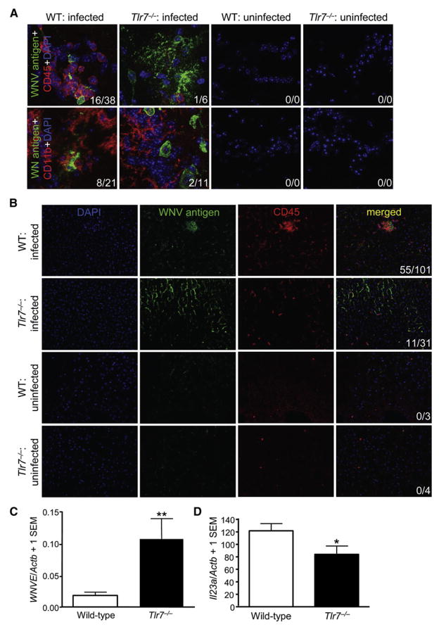
Figure 3. In Vivo Leukocyte Homing to West Nile-Infected Cells Is Tlr7 Dependent.
Wild-type (WT) or Tlr7−/− mice were i.p. challenged with WNV (LD50). Uninfected WT and Tlr7−/− mice were euthanized and processed side-by-side as negative controls.
(A) Perfused brains were isolated on day 6 postinfection, and WNV antigen (green signal) and CD45 (leukocyte common antigen, red signal) or CD11b (macrophage and microglia marker, red signal) were detected by immunofluorescence with a Zeiss ApoTome-equipped epifluorescence microscope (original magnification 63×).
(B) Perfused livers were isolated on day 3 postinfection, and WNV antigen (green signal) and CD45 (red signal) were imaged with a Zeiss Apo-Tome-equipped epifluorescence microscope (original magnification 20×). DAPI (blue signal) was used as a nuclear counterstain, and representative images are shown. Numbers of CD45+ or CD11b+ cells per image colocalized with WNV antigen+ areas (first number) and total CD45+ or CD11b+ cells per image (second number) are shown in the bottom right in (A) and (B).
(C) Quantitative PCR for WNVE in wild-type or Tlr7−/− livers at day 3 p.i. Graph shows means + 1 SEM.
(D) Quantitative PCR for Il23a in wild-type or Tlr7−/− livers at day 3 p.i. Graph shows means + 1 SEM. Similar results were observed in 2–4 independent experiments with at least n = 4 per group for each experiment. **p < 0.01 and *p < 0.05 compared to wild-type mice.

