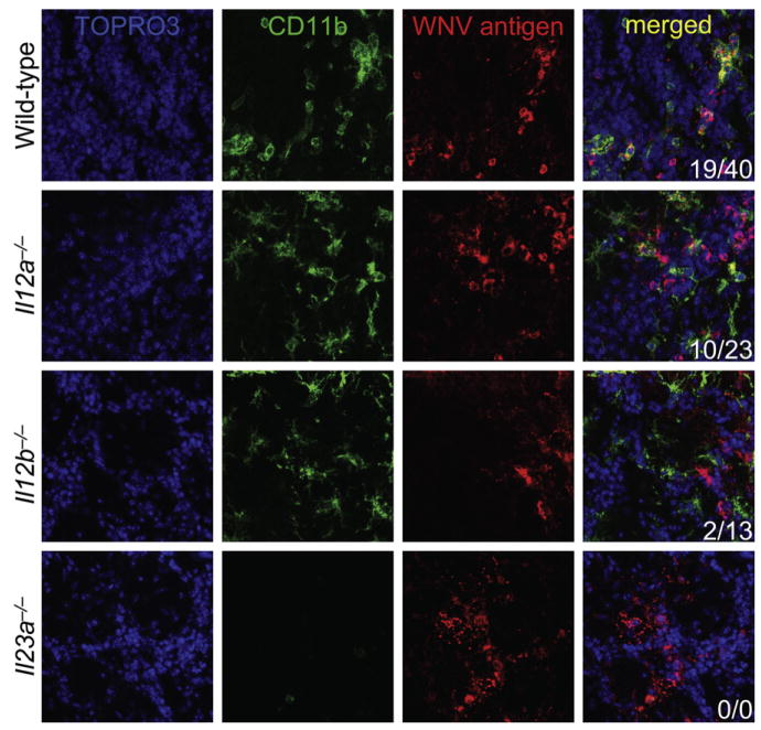
Figure 6. Macrophage Homing to West Nile Virus Is IL-23 Signaling Dependent.
Wild-type (n = 6), Il12a−/− (n = 4), Il12b−/− (n = 4), and Il23a−/− (n = 3) mice were infected with West Nile virus (LD50). Brains were isolated on day 6 after infection and immunostained for confocal microscopy with antibodies against CD11b (green signal) and WNV antigen (red signal) to reveal microglia and infiltrating macrophages in WNV-infected brain regions. TOPRO3 was used as a nuclear counterstain (blue signal) and merged images are shown to the right. Numbers of CD11b+ cells per image colocalized with WNV antigen+ areas (first number) and total CD11b+ cells per image (second number) are shown in the bottom right. Similar results were obtained in 2–4 independent experiments.

