Fig. 7.
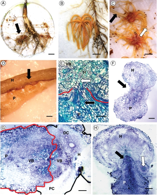
(A) Rhizotron-propagated root system of a sea carrot infected with O. minor ‘spiders’ (arrow); (B) ‘spider’ of O. minor var. minor on clover root; (C) ‘spiders’ of O. minor ssp. maritima on carrot roots (note the host root is distended before the point of parasite attachment, indicated by a black arrow, relative to post-attachment); (D) secondary haustoria of O. minor subsp. maritima parasite (P) attached to host root (H); (E) haustorial interface of O. minor on clover root with host (H) xylem elements (white arrow) and a concentration of parasite (P) xylem elements (black arrow) resulting in vascular connectivity (red lines mark the host–parasite interface); (F) haustorium of O. minor var. minor (P) attached to clover root (H) with vascular connectivity (arrow) at host–parasite interface; (G) O. minor ssp. maritima haustorium (P) attached to carrot root (H) showing connectivity of vascular bundles (VB) and massive distension of distal host cortical parenchyma (DC) relative to proximal host cortical cells (PC). Parasite tissue is marked in red, host tissue is marked in black; (H) O. minor var. minor haustorium (P) attached to host carrot root (H) with parasitic endophyte (black arrow) and secondary parasite vascular tissue differentiation. Scale bars: (A) 1 cm; (B) 4 mm; (C) 3 mm; (D) 0·5 mm; (E) 50 µm; (F) 200 µm; (G) 200 µm; (H) 200 µm.

