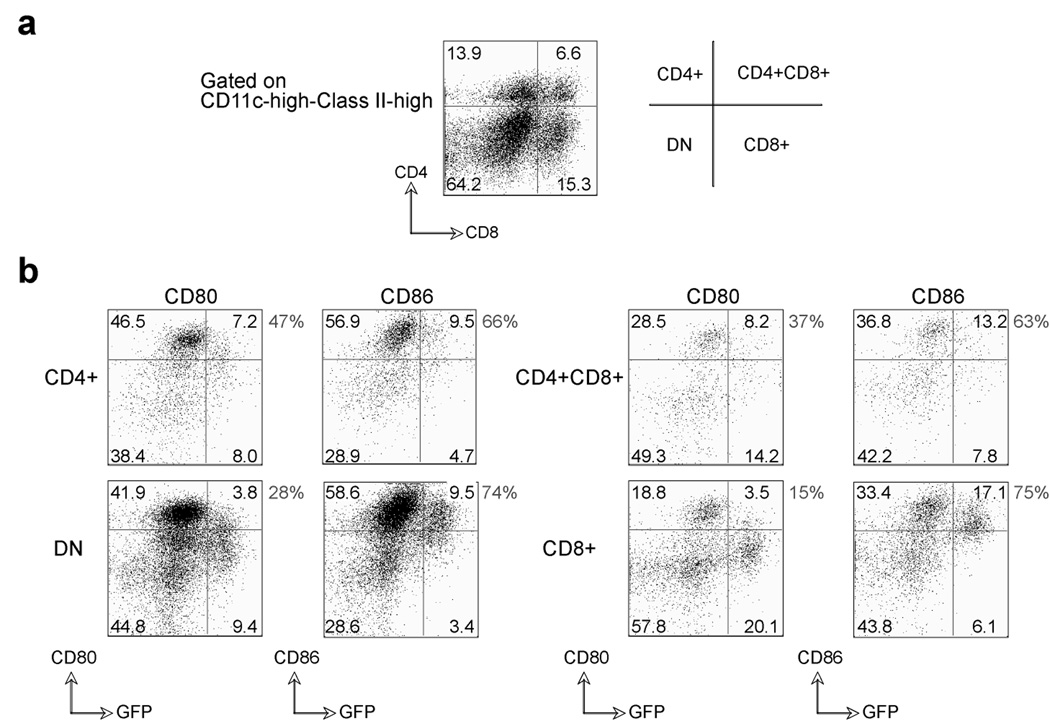
Figure 10. Differential expression of B7.1 and B7.2 on VACV-infected dendritic cell subsets.
Total splenocytes from BL6 mice were infected in vitro with VACV-GFP (MOI=10) for 24 hrs. (a) Representative plots of CD4 and CD8 staining, gating on CD11c-high/MHC Class-II-high cells. Percentages of DC subsets (CD4+, CD8α+, CD4+CD8α+, and double negative (DN)) in the spleen are indicated. (b) Upregulation of B7.1 and B7.2 was assessed on GFP positive and GFP negative DC subsets (CD11c-high, MHC Class-II-high). Percentages of DC subsets (CD4+, CD8α+, CD4+CD8α+, and double negative (DN)) positive for B7.1 and B7.2 are indicated within the plots, and percentages of GFP+ DC expressing B7.1 vs. B7.2 are shown outside the plots.

