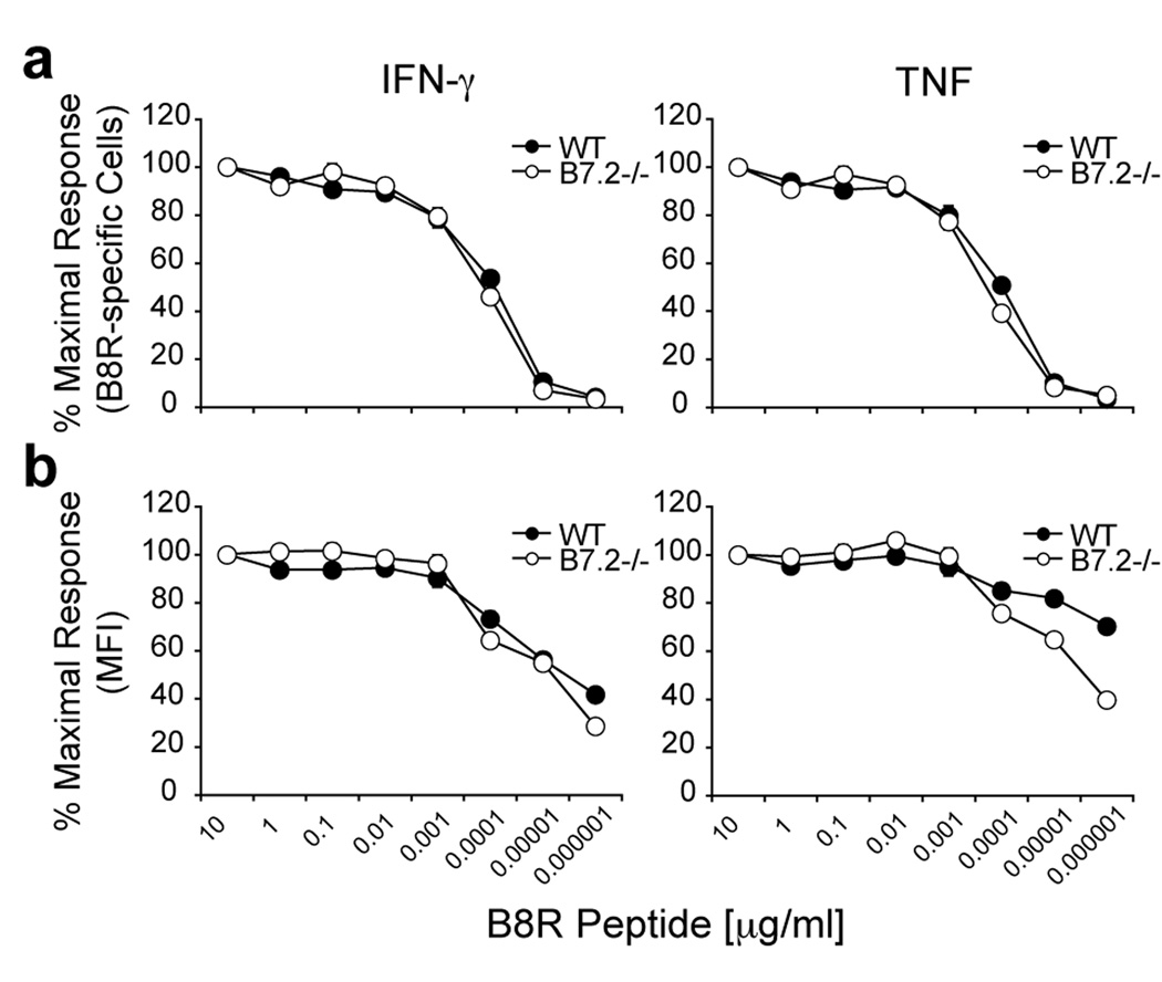
Figure 5. Differential reactivity of B8R-specific CD8 T cells in VACV-WR infected B7.2-deficient and wild-type mice.
Groups of C57BL/6 wild type or B7.2-deficient (B7.2 −/−) mice were infected i.p with VACV-WR (2 × 10⁵ PFU/mouse). Eight days post-infection splenocytes were harvested and stimulated for 6 h with graded concentrations of B8R peptide as indicated. (a) CD8 T cell reactivity was assessed by intracellular IFN-γ and TNF staining or (b) mean fluorescent intensity (MFI) on a per cell basis. The percentages or MFI of cytokine positive B8R-specific CD8 T cells were plotted against the peptide concentration used to stimulate the cells. Results are mean number ± SEM (n=4 mice/group) from one experiment. *, p < 0.05 (wt mice vs knockout) as determined by Student’s t test.

