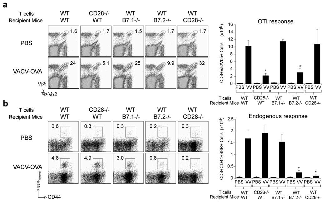
Figure 8. CD28 is required directly by CD8 T cells responding to VACV.
Naive WT or CD28−/− OT-I CD8 T cells were adoptively transferred into naïve WT B6, or B7.1−/−, B7.2−/−, or CD28−/− mice. One day later, mice were infected i.p. with recombinant VACV expressing full-length OVA (VACV-OVA; 2 × 10⁶ PFU/mouse) or PBS as indicated. (a) After 8 days, OT-I CD8 T cell expansion was analyzed by tracking the transgenic TCR. Dot plots: Representative co-staining for Vα2 and Vβ5 after gating on CD8 cells. Percent positive indicated. Right: Total numbers of CD8+Vα2+Vβ5+ cells per spleen. (b) Endogenous B8R-specific CD8 response after OT-I cell transfer. Naive WT or CD28−/− OT-I CD8 T cells were adoptively transferred into naïve WT B6, or B7.1−/−, B7.2−/−, or CD28−/− mice and infected with VACV-OVA as in (a). Seven days post-infection splenocytes were harvested and stained for CD8, CD44, B8R-tetramer. Left: Representative plots of tetramer staining, gating on CD8 cells. Percentages of activated B8R-tetramer positive CD8 T cells (CD8+CD44+B8R+) are indicated. Right: Total numbers of B8R-tetramer positive CD8+CD44+ T cells per spleen. Results are mean number ± SEM (n=4 mice/group) from one experiment. *, p < 0.05 (wt mice vs knockout) as determined by Student’s t test. Similar results were obtained in 1 additional experiment.

